Breast Density


About
WHAT IS BREAST DENSITY?
Your breasts consist of fat and fibroglandular tissues, which includes the glands and ducts
that support breastfeeding. Breast density is the amount of fibroglandular tissue compared
to your breast size.
Breast density is determined from your mammogram and not by the
way your breasts feel, or their appearance.
Risks
Dense breast tissue can mask cancer in mammograms which is why your clinician may recommend additional screening such as ultrasound.
Some studies say that the risk of breast cancer is six times greater for women with dense breast compared to low density breasts.
If you have dense breasts
High breast density is a normal condition, but it may affect the ability of the radiologist to find cancers on the mammogram, particularly when they are very small (and more treatable).
Supplementary screening (such as an ultrasound) can substantially improve cancer detection in women with high breast density and together with your clinician you can decide what the right steps are for you.
If your breasts are not dense, other factors may still place you at increased risk of breast cancer - including family history, and you should still get an annual mammogram starting at 40.

Will you still need a mammogram if you have dense breast?
Yes
There are certain tissue characteristics, like microcalcifications, that are imaged very well by mammography, but poorly or not at all by other imaging methods.

Case Report
HISTORY
A 50-year-old asymptomatic woman presented for routine screening in a private breast imaging facility.
Her mammogram and breast ultrasound 2 years ago showed no evidence of breast cancer.
Importance of screening
Screening mammograms have been proven to reduce breast cancer deaths so keep having them.
Fatty tissue is transparent to x-rays and appears near black on a mammogram. Dense tissue (and cancer) stops x-rays and appears white.
Breast density is classified into four categories by radiologist. Your technician will be able to tell you what your breast density is after your mammogram.
Our clinic uses Volpara, a volumetric breast density measurement tool, to better assess your breast density and are able to offer you a more personalised management approach of your breast health based on your risk factors.
Why is Breast Density Important?
Every woman has some amount of breast density. It is important to know your breast
density for managing your health, just like knowing your cholesterol score. Having high
breast density, like strong family history of breast cancer, has been linked to a higher risk of developing breast cancer. At Women’s and Breast Imaging, we use Volpara TruDensity™
software to automatically and objectively determine your breast density and provide you
with more accurate result
Image Findings

Figure 1. Bilateral craniocaudal (CC) and mediolateral oblique (MLO) mammograms show extremely dense breasts with more than 75% fibroglandular tissue (Volpara Score d)

Figure 2. (A) On ultrasound examination, there is an irregular solid mass measuring 12mm with suspicious sonographic features in the right breast 12 o’clock position. This mass was not palpable and obscured by the extreme density of the breast. (B) The biopsy procedure was done under ultrasound guidance using a standard 14-gauge biopsy needle.
This lump would grow in her right breast and remain undetected in her mammogram study for many scans without the ultrasound examination.
Pathology result confirmed a malignant pattern of an invasive lobular carcinoma, ER and PR positive and HER2 negative. She underwent mastectomy, axillary clearance and reconstructive surgery.
Discussion
The breasts are comprised of a combination of glands, ducts, fat and fibrous connective tissue. Fibroglandular breast tissue is white or bright on a mammogram. Fatty tissue is grey or dark on the mammogram (Figure 3). Breast density is a description of the relative amount of fibroglandular tissue vs fatty tissue. If the mammogram is more than 50% dense or white, it is then described as dense breast. Fatty or non-dense breast tissue is dark or grey on a mammogram with less than 50% whiteness/brightness. Cancerous tissue also appears white or bright on the mammogram which makes it very difficult for breast radiologists to detect any abnormality in a dense breast mammogram.

Figure 3. Normal Breast.
A. The normal breast is composed of milk-producing glands at the ends of ducts leading to the nipple. There is a layer of fat just beneath the skin, and often a few lymph nodes are seen near the underarm (axilla). B. On a mammogram, fat appears dark or gray; glandular tissue, fibrous tissue, muscle, and lymph nodes appear light gray or white. Masses due to cancer also appears white.
Source: Jeremy M. Berg. PhD. Breast Density: Why it Matters. DenseBreast-info, Inc. 2017
In Australia, breast density is typically described by visual assessment using BIRADS lexicon and can be classified into four categories by radiologist:
-
Less than 25% fibroglandular density = almost entirely fatty(Figure 4)
-
26-50% fibroglandular density = scattered fibroglandular areas (Figure 5)
-
51-75% fibroglandular density = heterogeneously dense (Figure 6)
-
More than 75% fibroglandular = extremely dense (Figure 7)

Figure 4. The breasts are almost entirely fatty (Volpara a) and almost completely grey or dark on mammogram examination.

Figure 5. The breasts are comprised of scattered fibroglandular areas (Volpara b) and 26-50% white or bright on mammogram examination.

Figure 6. The breasts are composed of heterogeneously dense (Volapra c) and 51-75% white or bright on mammogram examination.

Figure 7. The breasts are composed of extremely dense fibroglandular breast tissue and are more than 75% white or bright on mammogram examination.

Figure 8. This image demonstrates the different patterns of women’s breast density from fatty (top left) to completely white-out breasts (lower bottom). Visual designation of breast density may be subjective, inconsistent and not reproducible due to inter-observer and intra-reader variability.
.png)
Figure 9. Volpara is a volumetric breast density measurement tool that supplements the radiologist’s evaluation of mammogram studies. Volpara software is used locally and internationally to help radiologists assess breast density more objectively. It is an important tool to assist breast radiologists triage patients who will need an ultrasound to supplement their breast cancer screening.

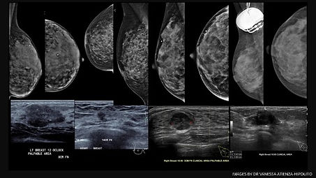
Figure 10. Companion case. This is an example of a fatty replaced left breast tissue with multiple biopsy-proven breast cancerous lesions and FNA-proven metastatic axillary lymph nodes well detected in mammogram and ultrasound examination.

Figure 11. Companion cases. These are examples of 4 patients with extremely dense breast tissue with biopsy-proven breast cancers only evident on ultrasound. These lesions are mammographically obscured on conventional 2D screening mammogram examination.


